LOCALITE is a frameless neuronavigation system that particularly addresses a problem with current interventional magnetic resonance imaging (iMRI) systems: non-interactive response time and poor image quality in the interactive mode. In addition, LOCALITE embeds iMRI images in a 3D enhanced-reality scenario that provides - in realtime - the cues a surgeon needs for effective spatial orientation during minimally invasive neurosurgery. The LOCALITE system lets the surgeon perform highly critical interventions more accurately and thus substantially reduces the risk for the patient.
LOCALITE is an off-spring of the VEP project. The project started in 1999 and was successfully completed with the CE-certification of the LOCALITE guiding system in May 2000. The LOCALITE guiding system is now in routine application for brain surgery at the Klinikum Krefeld, Germany.
A spin-off company LOCALITE GmbH, Bonn (Germany) has been founded that will further develop and market the LOCALITE system.
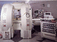
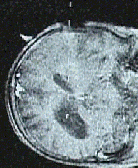
Interventional Magnetic Resonance Imaging (iMRI) systems [Figure 1] are designed to enable frameless minimally invasive surgery on targets that may move during the intervention (e. g. soft tissues). An interactive locator device tracks the surgical device. Slices of the body at the current position of the device are imaged to let the surgeon control the progress of the intervention. In interactive mode, however, iMRI systems generate only single 2D planes (real-time slices) [Figure 2] which do not give the surgeon sufficient context and orientation for finding efficiently the planned access path and the target. Each real-time slice is displayed with a delay of about 5 seconds. This prohibits interactive work. And the image quality is inadequate for the recognition of critical details.
 |
 |
|
|
|
To compensate for the deficiencies in image quality and response latency, LOCALITE uses simulated slices calculated from a volume data set generated intra-operatively by the iMRI system in scan mode. From this volume data set, any slice selected by the locator can be generated instantly and with high image quality.
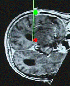
To compensate for the lack of context and orientation cues in the interventional MR images, LOCALITE embeds the simulated slice in a 3D enhanced-reality scene. During a planning phase, the position of entry and target are determined and coloured markers are generated at those positions [Figure 3].
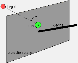
This scene does not yet provide sufficient cues for navigating the surgical device to the target area. Therefore, LOCALITE offers an additional abstract view [Figure 4] that shows the device in proper perspective relative to entry point and target. This navigation view supports hand-eye-coordination and efficiently guides the surgeon in positioning and maneuvring the surgical device.
 |
 |
|
Simulated slice with markers for entry and target Marking of the target, MPEG movie (1.8 MB) Control of the result, MPEG movie (0.7 MB) |
Abstract guiding scene supporting navigation Positioning the surgical device, MPEG movie (1.5 MB) Control of access path, MPEG movie (1.9 MB) |
Although a volume scan takes some 10 minutes, the volume is sufficiently close to the current situation such that the simulated slices caloculated from this volume can be used instead of the real-time images in the interactive mode in most cases. By this approach, the deficiencies of the real-time images are overcome: slices displayed with a delay of about 5 seconds and an image quality inadequate for the recognition of critical details.
Comparison of real-time and simulated slices with respect to timing and quality, MPEG movie (1.7 MB)
Before any critical decision, however, the surgeon will compare the current real-time slice with the simulated slice [Figures 2 + 3]. If both images no longer match, the simulated slice is updated by rescanning a volume intra-operatively.
PC with graphic card supporting OpenGL, attached to an iMRI system SIGNA SP from GE Medical Systems equipped with a FlashPoint 5000 locator device from Image Guided Technologies, Inc.
Uwe Behrens, Martin Bublat, Harald Busse, ernoth Grunst, Marko Jahnke, Matthias Jungmann, Klaus Kansy, Ralf Ratering, Arno Schmitgen, Sascha van Stockum, Peter Wisskirchen, Marc Wengler
Please contact: >Klaus Kansy, Gernoth Grunst, FhG-FIT